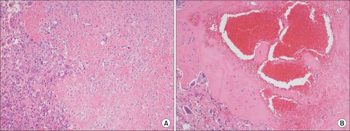
Fig. 2.

Sheets of tumor cells were intermingled with fine eosinophilic chondroid matrix (H&E, ×100). The tumor cells showed oval shaped nuclei with moderate eosinophilic cytoplasm. Several multinucleated giant cells and occasional mitotic figures were identified (A). Blood filled cystic cavities were consistent with secondary aneurysmal bone cyst (B).
