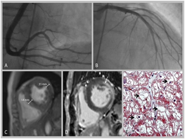
Figure 2. Cardiac work up in patient 2.
A+B: Coronary angiography (A: right coronary artery; B: left coronary artery) demonstrating no relevant pathology. C+D: Cardiac MRI showing increase in myocardial wall thickness (C) and pathological late gadolinium enhancement (D, arrow). E: Myocardial biopsy revealing strong accumulation of Gb3, as indicated by numerous vacuoles within the cardiomyocytes (arrow).

