
Figure 1.
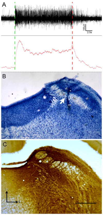
Parabrachial nucleus injection site is in gustatory responsive waist region. (A) The coordinates of gustatory responsive PBN region were determined in initial in vivo recording experiments. Both multiunit (top trace) and integrated activity (bottom trace) display a sustained increase in neural activity throughout the duration of tastant application to the oral cavity (Stimulation start at green dashed line, rinse at red dashed line). (B) Nissl staining of the parabrachial nucleus in one case that received triple injections reveals distinct and local damage to the PBN waist region after BDA injection via Hamilton syringe. Injection not centered on the waist region failed to produce retrograde cellular filling in the rNTS despite diffusion of tracer away from the injection location into the waist region. Arrow marks the location of the Hamilton syringe tract passing through the waist region of the PBN. (C) An ABC-DAB labeled section from another case with triple injections illustrates the dense tracer labeling centered at waist region. D = dorsal, L = lateral. PBN subregions: w = waist region, bc = brachium conjunctivum, el = external lateral, m = medial, vl = ventral lateral. Scale in B = 600 μm.