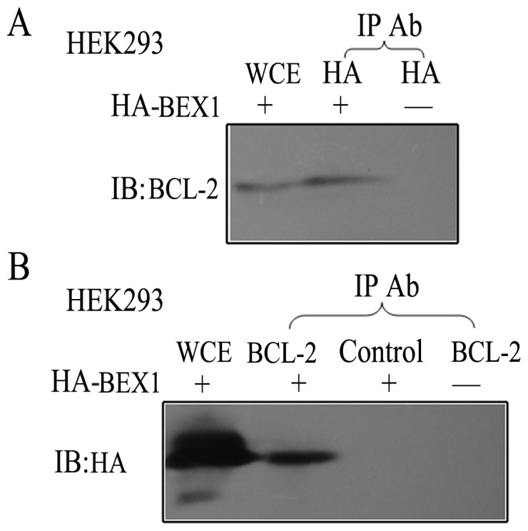
Figure 1. BEX1 interacts with the anti-apoptotic protein BCL-2.

“+” and “−” refers to whether or not cells were transfected with HA-BEX1. If the cells were not transfected with HA-BEX1, an empty vector pCMV-HA was used for transfection. “Control” refers to the rabbit IgG isotype control. A, BEX1 was immunoprecipitated using an anti-HA antibody, and the co-immunoprecipitate (co-IP) was analyzed by immunoblotting (IB) with an anti-BCL-2 antibody. The intrinsic BCL-2 was monitored in the whole-cell extract (WCE). B, BCL-2 was immunoprecipitated using an anti-BCL-2 antibody or isotype control rabbit IgG, and the co-IP was analyzed by IB with an anti-HA antibody. The presence of HA-BEX1 was also monitored in the WCE. The lower band was a non-specific signal.
