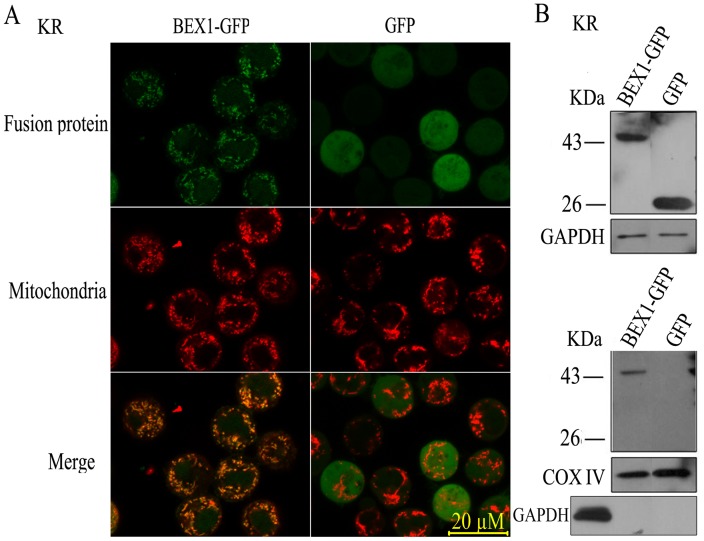
Figure 2. BEX1 localizes to the mitochondria.
A, Fluorescence of live KR cells expressing BEX1-GFP or an empty vector control (GFP). Cells were visualized for GFP (top), Mitotracker (middle), or merged images (bottom). B, Biochemical fractionation. WCE prepared from KR cells expressing BEX1-GFP or an empty vector control (GFP) were separated into cytoplasmic (top) and mitochondrial (bottom) fractions and then immunoblotted for GFP, GAPDH (a cytoplasmic protein), or cyclooxygenase IV (COX IV, a mitochondria marker). Cross contamination of mitochondrial fractions in the cytoplasmic fractions was excluded by immunoblotting with GAPDH.

