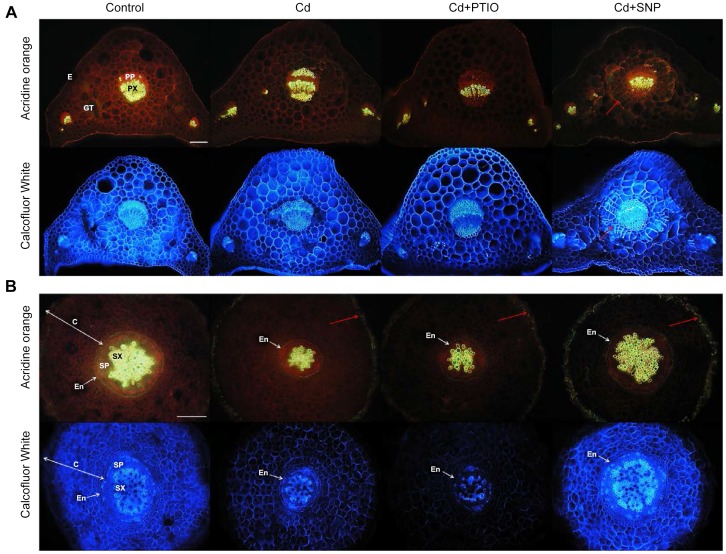
Figure 5. Changes in anatomy (transversal sections) of chamomile leaves (petioles, A) and roots (B) in hydroponically-cultured plants (using treatments as in the Figs. 1–4).
Acridine orange stains cellulose (red fluorescence), autofluorescence of lignin, cutin, and suberin remains unchanged. Calcofluor white stains cellulose (blue). See explanation for observed changes in the discussion. E – epidermis, GT – ground tissue (parenchyma), PP – primary phloem, PX – primary xylem, C – cortex, SP – secondary phloem, SX – secondary xylem, En – endodermis. Bar indicates 200 µm. Red arrow indicates parenchyma cells with increased amount of cellulose in cell walls (A) and suberinization of the outermost cells of cortex in all treatments with Cd (B).

