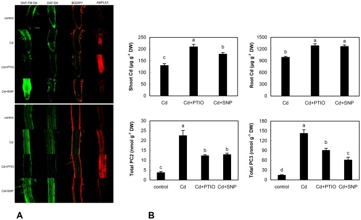
Figure 6. Confocal microscopy of NO changes (stained with DAF-DA and DAF-FM DA), lipid peroxidation (stained with BODIPY 581/591 C11) and general indicator of H2O2 (Amplex UltraRed) in root tips (upper panel) and upper part of roots (lower panel) of chamomile seedlings cultured on Petri dishes over 48 h with identical concentrations of Cd2+, PTIO and SNP as mentioned in Fig. 1.
Bar indicates 100 µm (A). Changes to content of Cd and phytochelatins (PC2 and PC3) in these seedlings (B). Total PC means that whole seedlings (shoot+root) were extracted.

