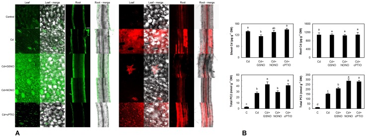
Figure 7. Confocal microscopy of NO changes (stained with DAF-FM DA, green emission) and general indicator of H2O2 (Amplex UltraRed, red emission) on the surface of cotyledons (leaf, first column in each panel) and in upper part of roots (third column in each panel) of chamomile seedlings cultured on Petri dishes with alternative NO modulators (S-nitrosoglutathione/GSNO - 300 µM, diethylamine NONOate/NONO - 300 µM and 2-(4-carboxyphenyl)-4,4,5,5-tetramethylimidazoline-1-oxyl-3-oxide/cPTIO - 60 µM) and 60 µM Cd2+ over 48 h (A).
Bar indicates 25 µm for leaf and 50 µm for root. Changes to content of Cd and phytochelatins (PC2 and PC3) in these seedlings (B). Total PC means that whole seedlings (shoot+root) were extracted.

