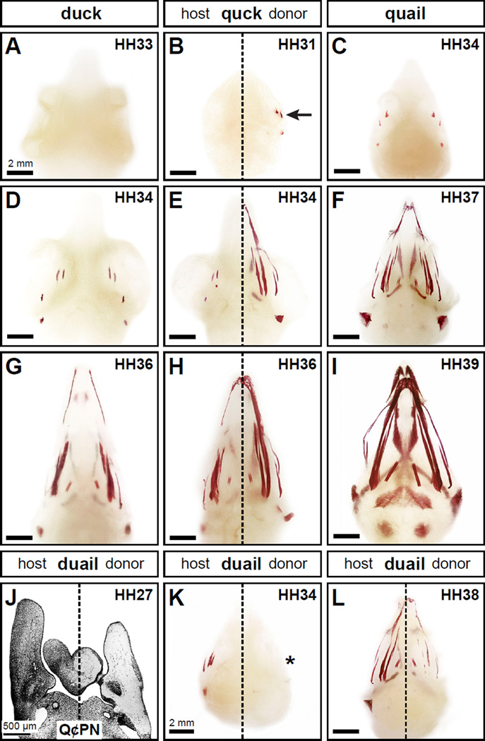
Figure 2. Neural crest mesenchyme controls the timing of mineralization.
(A) Neither duck nor quail (not shown) display any signs of mineralization in the head skeleton at HH33 following Alizarin red staining as shown in ventral view. (B) In chimeric quck at HH31 (n=4), there is no sign of mineralization on the duck host side but the quail donor side has begun to mineralize (arrow) like that observed in quail three stages later. (C, D) Craniofacial mineralization in quail and duck can first be detected at HH34. (E) The pattern of mineralization on the quail donor side in quck at HH34 (n=8) is similar to that found in quail at HH37 (F) . (G) Similarly, mineralization in duck at HH36 is like the host side of quck at HH36 (H) whereas the donor side resembles that of quail at HH39 (I) (n=8). (J) In reciprocal transplants that generate chimeric duail, the host side is labeled with Q¢PN-positive quail cells (black) whereas the duck donor side is unlabeled as shown in a coronal section through the mandible at HH27. (K) In the heads of chimeric duail stained with Alizarin red, the quail host side follows its normal time course for development and is mineralized at HH34 (n=3). In contrast, on the duck host side mineralization is delayed (asterisk). (L) By HH38, the duck donor side of chimeric duail has begun to mineralize (n=3).

