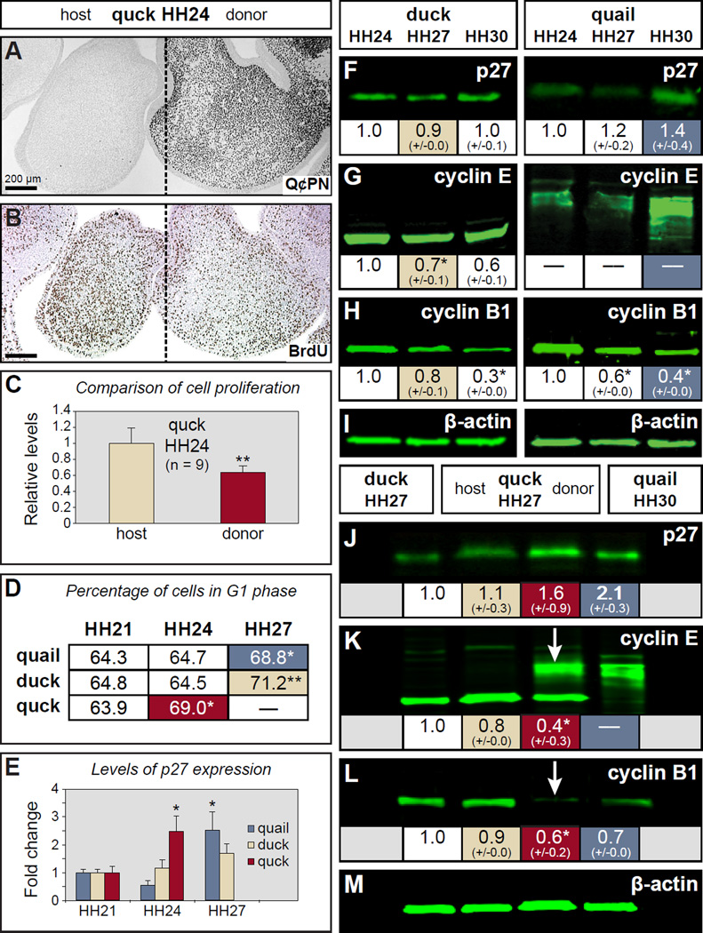
Figure 6. Neural crest mesenchyme regulates cell cycle progression.
(A) Section of an HH24 quck showing Q¢PN-positive quail donor NCM (black cells) on one side of the mandible as well as unlabeled duck host tissues. (B) An adjacent section stained with an antibody to BrdU (dark brown) showing a decrease in the amount of proliferating cells coincident with the distribution of faster-developing quail donor NCM. (C) Quantification of BrdU-positive cells in HH24 quck demonstrates a significant decrease in proliferation on the quail donor side compared to the duck host side. (D) At HH24, donor NCM in mandibles from chimeric quck contains a significantly higher percentage of cells in G1 arrest (red-shaded box) versus cells cycling through G2, M, and S phases, which is more like that observed in quail (blue-shaded box) and duck (beige-shaded box) at HH27, as determined by FACS. (E) RT-qPCR reveals that the level of p27 mRNA in quck at HH24 is significantly more like that observed in quail at HH27. For panels C , D , and E , the quantitative data represent the mean ± SEM. *, p≤0.05; **, p≤0.01. (F-I) Western blots showing p27, cyclin E, and cyclin B1 expression levels in quail and duck at HH24, HH27, and HH30. β-actin was used as a loading control. (J-M) Western blots of host and donor sides of quck at HH27 compared to control HH27 duck and HH30 quail reveal that quail donor NCM express species and stage-specific patterns of protein expression, which can be clearly seen as quail-like for cyclin E (K, arrow ) and cyclin B1 (L, arrow ) on the donor side. For panels F-M , the images represent protein levels from a single run of the samples whereas the values shown below are the means of quantified protein levels from multiple runs of the samples ± SEM. * p≤0.05.

