
Fig. 2.
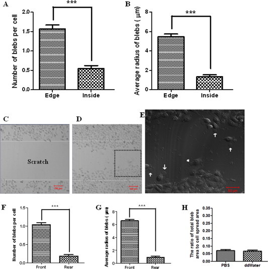
Asymmetric distribution of fixation-induced blebs on spread or migrating HUVECs. Cells were fixed with 4% paraformaldehyde at room temperature for 20 min. (A) The average number of blebs per cell and (B) the average radius of blebs distributing at the edge/boundary or at the inside/body of spread cells. (C) A “scratch” field was shown before cell migration. (D) Many cells migrated into the “scratch” field. (E) The higher-magnification image of fixed migrating cells in the area enlarged from the dashed square in Fig. 2D. Some blebs at the front (arrows) and rear (arrowhead) edges were indicated. (F) The average number of blebs per cell and (G) the average radius of blebs distributing at the front or rear edge of migrating cells. (H) Slight changes in ionic strength and pH value of fixative solutions have no significant effects on the genesis of blebs. The terms “PBS” and “ddWater” in the graph means the cells fixed by 3% paraformaldehyde (pH ∼7.2) and 3% paraformaldehyde (pH ∼7.3), respectively. The fixative solutions were prepared by diluting 4% paraformaldehyde with PBS and double distilled water (ddWater), respectively, causing slight differences in ionic strength and pH value of the two fixative solutions. Scale bar: (C, D) 100 μm; (E) 50 μm.