
Fig. 6.
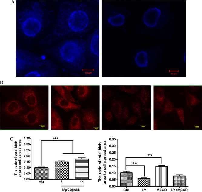
Effect of MβCD on fixation-induced blebbing of spread HUVECs. (A) MβCD causes the loss of cholesterol in the plasma membrane of HUVECs. HUVECs were pre-treated with (right) or without (left) 10 mM MβCD and then subjected to filipin staining and confocal microscopy. Scale bar: 10 μm. (B) MβCD causes the loss of PIP2 in the plasma membrane of HUVECs visualized by confocal microscopy. From left to right: no treatment; treated with 50 μM LY294002 for 30 min; treated with 10 mM MβCD for 30 min; treated with LY294002 first and then MβCD. Scale bar: 10 μm. (C) Quantitative analyses of fixation-induced blebs on MβCD-treated HUVECs. Left panel: HUVECs were pre-treated with MβCD (0, 5, or 10 mM for 30 min) and then fixed with 4% paraformaldehyde at room temperature for 30 min. The number of cells measured in each group are indicated in Table 1 (∗∗∗p < 0.001 compared with the control). Right panel: the cells were treated as indicated in Fig. 6B (∗∗p < 0.01 compared with the control).