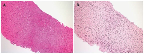
Figure 2.

Needle liver biopsy of male infected with human immunodeficiency virus. A: His risk factor for the development of Nodular Regenerative Hyperplasia was long term use of didanosine. Hepatocytes size varies zonally, occasional areas of small hepatocytes with increased nuclear cytoplasmic ratio alternate with areas of a more normal appearing morphology in the hematoxylin-eosin stain (original magnification × 100); B: The reticulin stain highlights and confirms nodular regeneration throughout the specimen with nodular areas of regenerative hepatocytes as characterized by widened hepatocellular plates alternating with areas of normal appearing lobular architecture and then areas of narrowed, attenuated hepatocytes (original magnification × 100).
