Fig. 1.
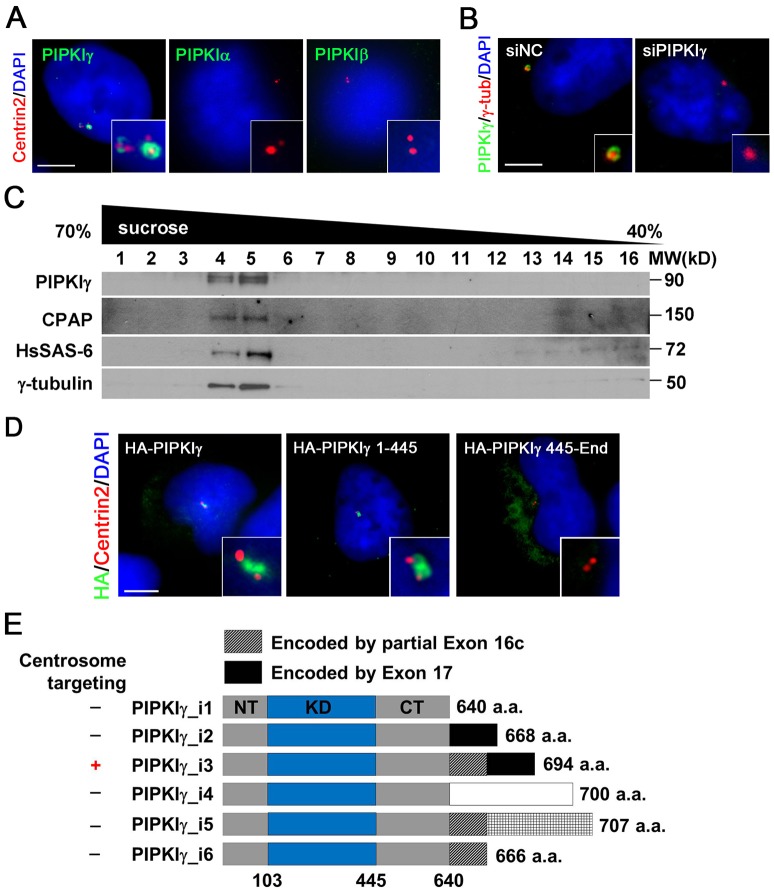
PIPKIγ associates with the centrosome. (A) PIPKIγ, but not PIPKIα or PIPKIβ, is targeted to the centrosome. HeLa cells were fixed and stained with the indicated antibodies to visualize PIPKIγ, PIPKIα or PIPKIβ (green) along with centrin 2 (red). (B) siRNA-mediated depletion of PIPKIγ abolishes the centrosome signal recognized by anti-PIPKIγ antibody. HeLa cells were treated with nonspecific control (siNC) or PIPKIγ-specific (siPIPKIγ) siRNA and then stained with the indicated antibodies. Anti-γ-tubulin was used to label centrosomes. (C) PIPKIγ co-sedi-ments with centrosomes in 40–70% sucrose gradients. Centrosome fractions were isolated from HeLa cells and immunoblotted using the indicated antibodies. (D) HeLa cells were transfected with the HA-tagged full-length (HA–PIPKIγ), N-terminal fragment (HA–PIPKIγ 1-445) and C-terminal fragment (HA–PIPKIγ 445-End) of PIPKIγ_i3. Cells were then stained with anti-HA (green) and anti-centrin 2 (red) antibodies. (E) Schematic diagram comparing the sequence and centrosome targeting of known PIPKIγ isoforms. NT, N-terminus; KD, kinase domain; CT, C-terminus. PIPKIγ_i3 is the only isoform localized to centrosomes. The white shading represents the polypeptide translated from exon 16b, the hatched shading represents the polypeptide translated from exon 16c of the pip5k1c gene. Scale bars: 5 µM (A,B,D). Enlarged centrosome images were shown as inserts. DNA was stained with DAPI.

