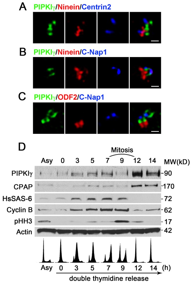
Fig. 2.

PIPKIγ localizes around the proximal end of centrioles and its protein level is regulated by the cell cycle. (A-C) HeLa cells were subjected to indirect immunofluorescence microscopy to visualize PIPKIγ along with ninein and centrin 2 (A), ninein/C-Nap-1 (B) and ODF2/C-Nap-1 (C) using 3D-SIM. SIM images of representative centrosomes show the positions of PIPKIγ and the indicated centrosome markers. These images indicate that PIPKIγ localizes below the distal and subdistal appendages and close to the proximal end of centrioles. Scale bars: 0.5 µm. (D) HeLa cells released from a double thymidine block were collected at the indicated time-points and were analyzed by immunoblotting using appropriate antibodies. The lower panel shows flow cytometry analyses of propidium iodide-stained cells at each time-point, confirming successful synchronization. Asy, asynchronous cells.
