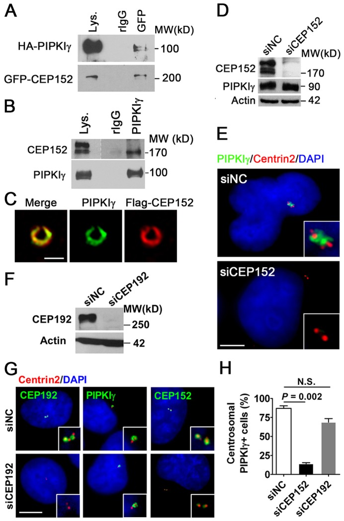
Fig. 3.

CEP152 associates with PIPKIγ and regulates PIPKIγ targeting to the centrosome. (A,B) CEP152 and PIPKIγ potentially associate with one another. (A) HEK293T cells co-transfected with vector encoding HA–PIPKIγ and GFP–CEP152 were subjected to immunoprecipitation using normal rabbit IgG (rIgG) or rabbit anti-GFP (GFP) antibodies. Lys., cell lysate. (B) Co-precipitation of endogenous PIPKIγ and endogenous CEP152 from HeLa cells. The precipitates from A and B were analyzed by immunoblotting with the indicated antibodies. (C) PIPKIγ and CEP152 colocalize at the centrosome. HeLa cells expressing FLAG–CEP152 were stained with anti-PIPKIγ and anti-FLAG antibodies and then analyzed by 3D-SIM. (D) CEP152-specific siRNA (siCEP152) efficiently depleted endogenous CEP152 from HeLa cells. siNC, non-specific control siRNA. (E) Loss of CEP152 eliminates endogenous PIPKIγ localization at centrosomes. HeLa cells were treated with siNC or siCEP152 and then stained with the indicated antibodies. Representative images of control or CEP152-depleted cells are shown. (F) CEP192-specific siRNA (siCEP192) efficiently depleted endogenous CEP192. (G) PIPKIγ localization at centrosomes was not significantly affected in CEP192-depleted cells, although CEP192 was completely abolished from the centrosome and the centrosomal signal of CEP152 was strongly decreased in these cells. (H) Quantification of the number of cells with robust centrosomal PIPKIγ staining in each group described in E and G indicates that the loss of CEP152, but not CEP192, significantly eliminates PIPKIγ targeting to the centrosome. Results from at least three independent experiments were analyzed, with >200 cells examined in each experiment. Error bars indicate s.d.; N.S., no significant difference. (E,G) Enlarged centrosome images are shown as inserts. Scale bars: 0.5 µM (C) 5 µM, (E,G). DNA was stained with DAPI.
