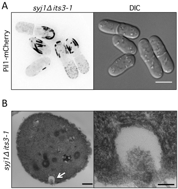
Fig. 4.

Pil1 generates cortical pits in syj1Δ its3-1 double-mutant cells. (A) Thick Pil1 filaments at the cortex of syj1Δ its3-1 double-mutant cells. Images are inverted maximum projections from Z-planes in the top half of the cell. Scale bar, 5 µm. (B) Thin section electron microscopy of syj1Δ its3-1 double-mutant cells. Cross-section view displays exaggerated pit-like invagination. The white arrow highlights a pit-like invagination that is magnified in right panel. Scale bars: 500 nm (left panel); 100 nm (right panel).
