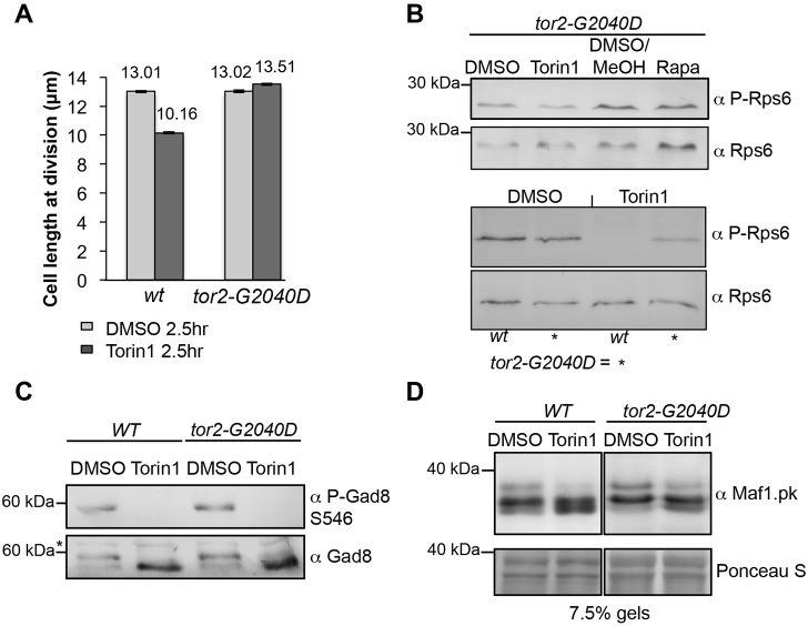
Fig. 4.
The tor2.G2040D mutation alters the dephosphorylation of TORC1 substrates following Torin1 treatment. (A) Cell length at division of indicated strains (n = 200). After 2.5 hours of Torin1 treatment, 10% of wt cells divide (see Fig. 5A). (B-D) Western blot using the indicated antibodies. Cells were treated as in Fig. 2. (C) Cells were treated with 25 µM Torin1 for 10 minutes. A non-specific band is indicated by an asterisk (*). (D) Cells were treated with 750 nM Torin1 for 10 minutes.

