
Figure 3.
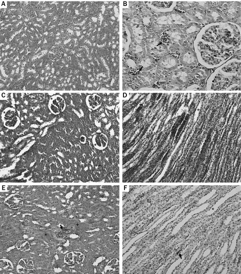
(A) Normal glomerular and tubular structure (hematoxylin and eosin [HE] ×100). (B) Neutrophil casts (arrow) (HE ×400). (C) Diffuse glomerular shrinkage (asterisk) (HE ×200). (D) Extensive vascular congestion, tubular degeneration, and dilatation (asterisk) (HE ×200). (E) Interstitial fibrosis (asterisk), cellular casts (arrow), hyalinization (plus sign), and degenerated glomeruli (HE ×200). (F) Mild tubular degeneration, dilatation (asterisk), and hemorrhage (arrow) (HE ×200).