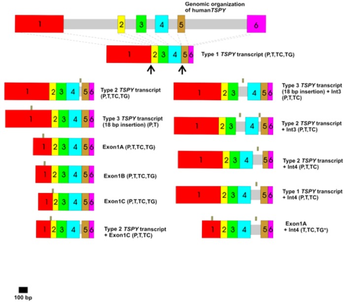
Figure 2.
Schematic illustration of a human TSPY gene and the various TSPY transcripts in human prostatic (P, prostatic adenoma and benign prostatic hyperplasia) and testicular (T, human testis; TC, human testicular seminoma) tissues [17] and in mouse testes of the transgenic line Tg(TSPY)9Jshm (TG) [47,50,51]. Introns are shown as grey bars and exons indicated as colored boxes. Small grey bars above the splice variants indicate the use of alternative splice donor and/or acceptor sites. The TSPY cDNA region encoding the cyclin B binding domain (SET/NAP domain) is highlighted by a black line (aa residues 121-265) [22]. * [52]. Detailed descriptions of the differential spliced TSPY transcripts are given in reference [17]. Splice variants are termed in accordance to references [17] and [26]. 18 bp insertion: in frame insertion of GTG GAG CTG GTG GCG CAG within exon 1 of human TSPY, +: combination of different splicing patterns.

