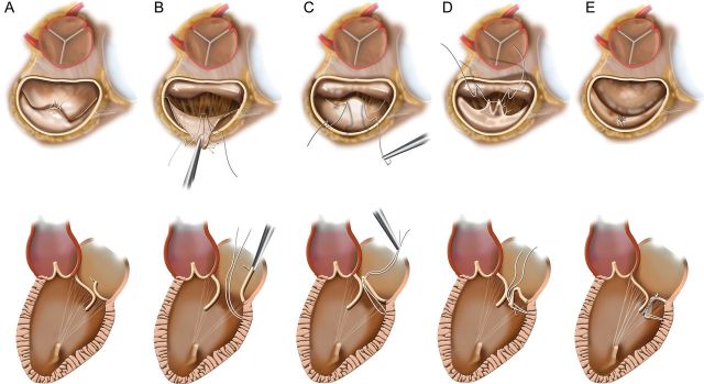
Figure 1:

(A) Short- and long-axis drawings depicting degenerative mitral disease with posterior leaflet ruptured chords and prolapse/flail. (B) The prolapsed segment is retracted into the atrium to expose the posterior ventricle just beneath the leaflet, and an anchoring CV5 Gore-Tex suture has been placed. (C) The suture has been loosely tied and the ends brought through the leading edge of the prolapsed segment. (D) The prolapsed segment edge is inverted into the left ventricle, generating two near-apposing radial folds that are then sutured together. (E) This eliminates the prolapse, yielding a smooth broad coaptation surface that is mobile yet positioned posteriorly enough to preclude anterior displacement and SAM.
