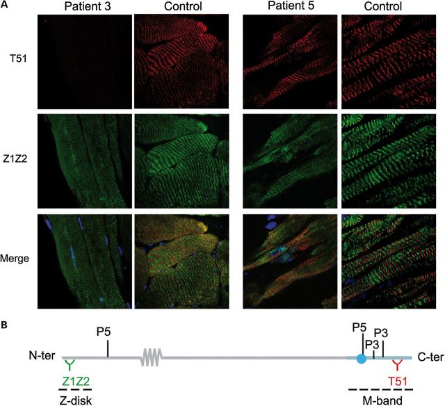
Figure 2.
Expression and sarcomere integration of mutant titin in Patients 3 and 5. (A) Quadriceps from P3 and control (left panel, 63×) and myocardium from P5 and control (right panel, 63 × 4); longitudinal cryosections. Note total loss of labeling with T51 in P3. Dapi (blue) stains nuclei. (B) Recognition sites of Z1Z2 and T51 anti-titin antibodies, respectively, upstream and downstream of P5 and P3 mutations.

