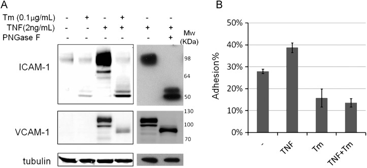
Fig. 1.
Tunicamycin prevented TNF-triggered ICAM-1 increase and monocyte attachment. (A) HUVECs were treated with dimethyl sulfoxide (DMSO) or 0.1 μg/mL tunicamycin for 24 h, and then treated with or w/o 2 ng/mL TNFα for 6 h. Total proteins were harvested and TNF-treated lysates were also treated with Peptide -N-Glycosidase F to remove the N-glycans. Tubulin is used as protein loading marker. (B) HUVECs grow into 100% confluence in 96-well plate followed by treating with DMSO or 0.2 μg/mL tunicamycin for 16 h, and then were treated with or w/o TNF (2 ng/mL) for 6 h. The Calcein labeled THP-1 cells were mounted to the HUVEC monolayer. The adhesion percentage is calculated as the fluorescence of adherent cells divided by the total fluorescence of cells added to well. Each error bar in the histogram represents standard deviation (SD) of three independent assays.

