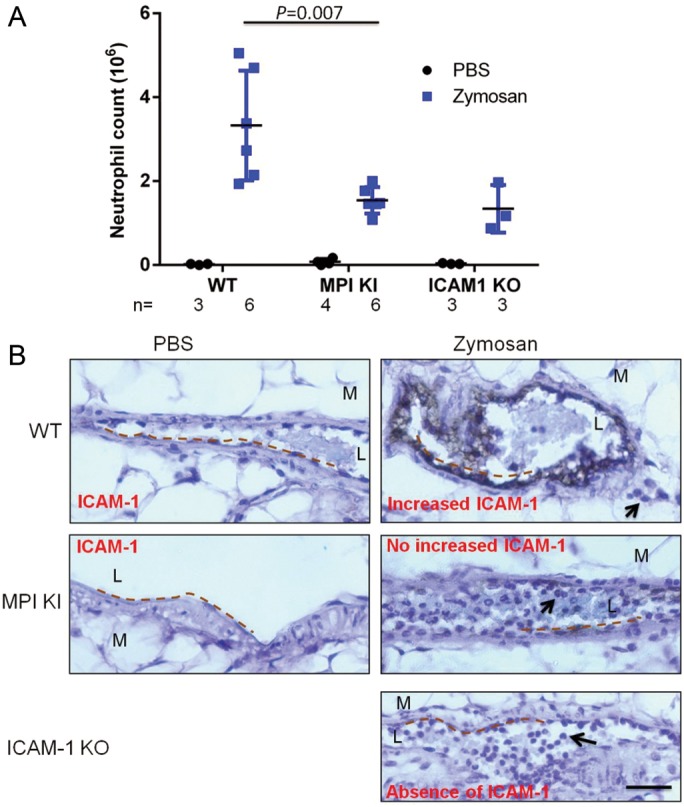
Fig. 3.

MPI deficient mice exhibited decrease of leukocytes extravasation in response to acute peritonitis. (A) Mice aged 6–10 weeks were i.p injected with zymosan or PBS, and the peritoneal lavage fluid was collected 4 h later. The Gr-1 positive cells by fluorescence-activated cell sorting analysis were defined as neutrophils. The total cell counts were calculated by TC20 automated cell counter. The absolute neutrophil number was calculated as total cell count × neutrophil%. (B) After zymosan injection for 4 h, the mesenteries were collected and fixed. The paraffinized slides were immunohistochemically stained by anti-ICAM-1 antibody, and ABC kit and DAB substrate were used to detect positive staining followed by hematoxylin counterstain. The images were captured with a Nikon Eclipse E800 microscope (Tokyo, Japan), using a Nikon ×40/0.75 air objective lens and a Nikon Digital Camera DXM2000. Images were processed using the Nikon ACT-1 (version 2.7) software and ImageJ 1.41 (NIH). The dotted lines indicate the vascular endothelial surface. The black arrows show the transendothelial leukocytes in wild-type and accumulated leukocytes within the vascular lumen in Mpi knockin (KI) and ICAM1 KO mice in acute inflammation. L, lumen; M, mesentery. The black bar represents 50 µm. Images were obtained from 1 of 3–6 individual mice, which gave representative results.
