
Figure 2.
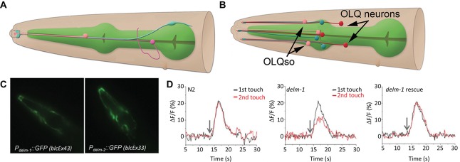
The amphid sheath and outer labial sensilla socket cells. (A) The amphid sheath glia (blue) cell body is positioned near the nerve ring (not shown) and sends a long, thin process, along with a neuronal dendrite (magenta) and the amphid socket cell process (pink) to the anterior tip of the worm (left side, dashed box); adapted from wormatlas.org. (B) A cartoon showing the outer labial sensilla sheath (OLLsh and OLQsh, blue) and socket (OLLso and OLQso, pink) cells and their extensions to the anterior of the worm where they ensheath the ciliated dendrite of the neurons (OLL and OLQ, red); note that inner labial sensilla is not shown here; adapted from wormatlas.org. (C) The promoters for the delm-1 and delm-2 drive reporter GFP expression in the outer and inner labial sensilla socket glial cells (OLQso and ILso, respectively) of worms (strains blcEx43 and blcEx33, respectively). (D) Knock out of delm-1 leads to reduced OLQ neuron calcium response to mechanical stimulation of the worm (middle), while re-expression of the channel in OLQso glia (using the glial promoter itx-1) rescued the neuronal responsiveness (right); N2, background strain (left). C and D adapted from Han et al. (2013).