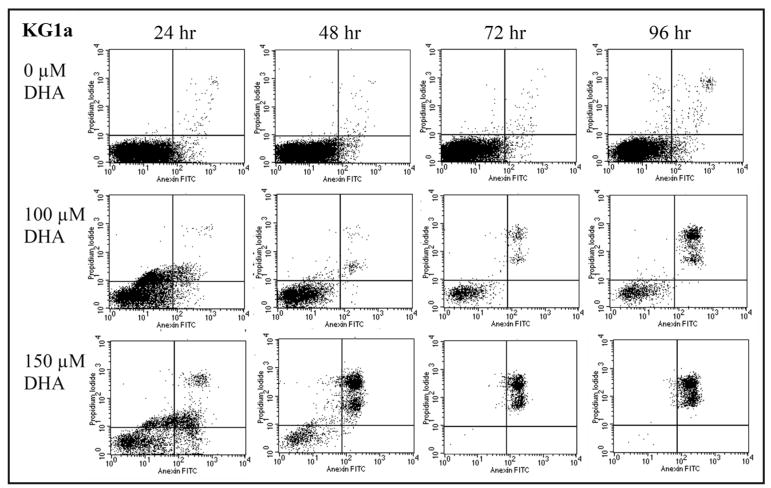
Figure 3.

DHA induces dose dependent apoptotic cell death in KG1a. KG1a cells cultured with 0, 100 or 150 μM DHA and stained with Annexin V and PI were analyzed daily by flow cytometry. Shown are the results from a representative experiment. Cells in early apoptosis are Annexin V positive/PI negative and appear in the lower right quadrant. Cell death is characterized by positivity for both Annexin V and PI and these cells appear in the upper right quadrant. While the untreated control had a very low percentage of cell death, KG1a cultures with 150 μM of DHA had cell death rates of 8.9, 30.4, 99.5 and 98.5%, respectively, at the four time points analyzed.
