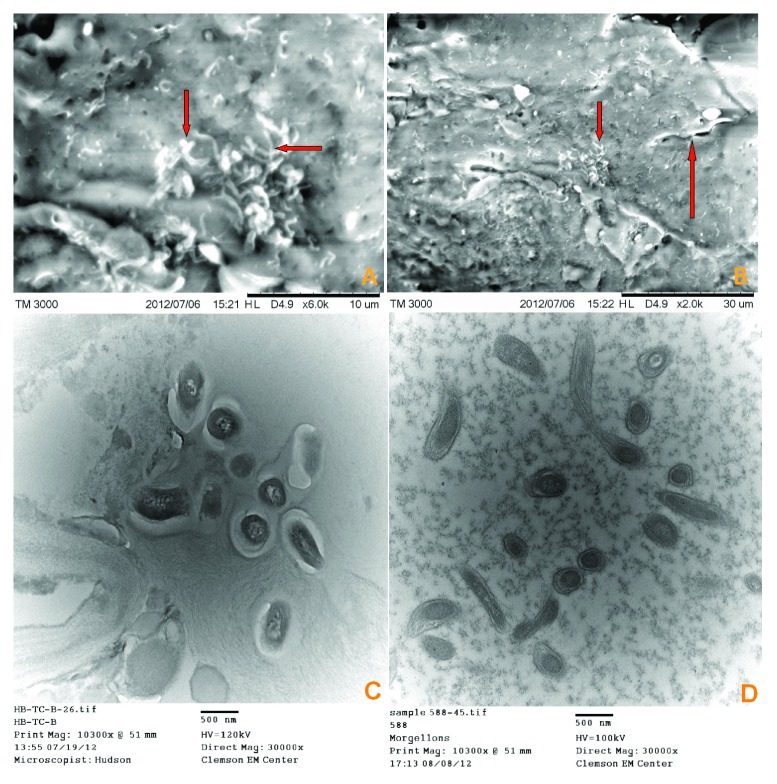
Figure 3.
A) SEM from patient 2 tissue sample showing spirochete images that are consistent with morphological forms of Borrelia (arrows). B) SEM from patient 2 tissue sample showing single spirochete, upper middle right (long arrow) and morphological forms consistent with Borrelia, center (short arrow). C) TEM from patient 1 tissue sample showing sectioned spirochetes. D) TEM from BDD tissue sample showing sectioned spirochetes.

