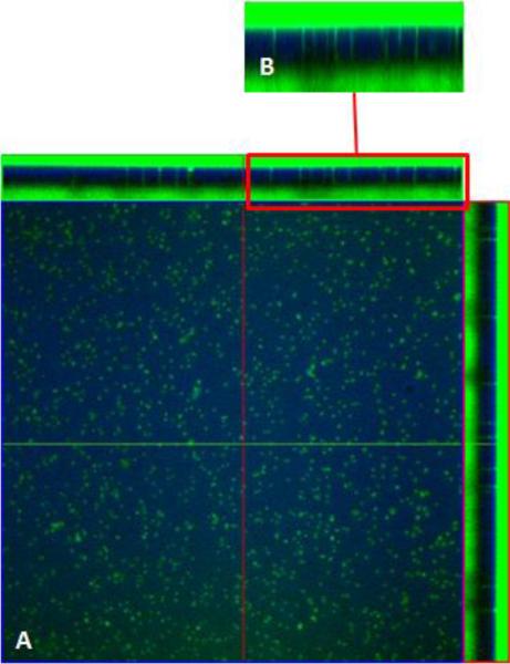
Figure 9.
Celltracker green was incubated on blank PET membranes as a positive control. Confocal images of cell tracker green localization on blank PET membrane (blue). The central portion of the panel is an en face view of the PET filter ,A, shown from the Z-axis, B(top)is a cross sectional view in the Z-plane of each panel; Celltracker green on the apical and basal side (green) of the PET membrane (blue). Note that pores are filled with Celltracker green.

