Figure 2.

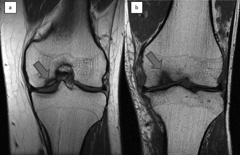
Coronal T1 weighted magnetic resonance imaging of the knee showing an area of osteochondritis dissecans affecting the medial femoral condyle (A) and an osteochondral lesion affecting the medial femoral condyle (B)

Coronal T1 weighted magnetic resonance imaging of the knee showing an area of osteochondritis dissecans affecting the medial femoral condyle (A) and an osteochondral lesion affecting the medial femoral condyle (B)