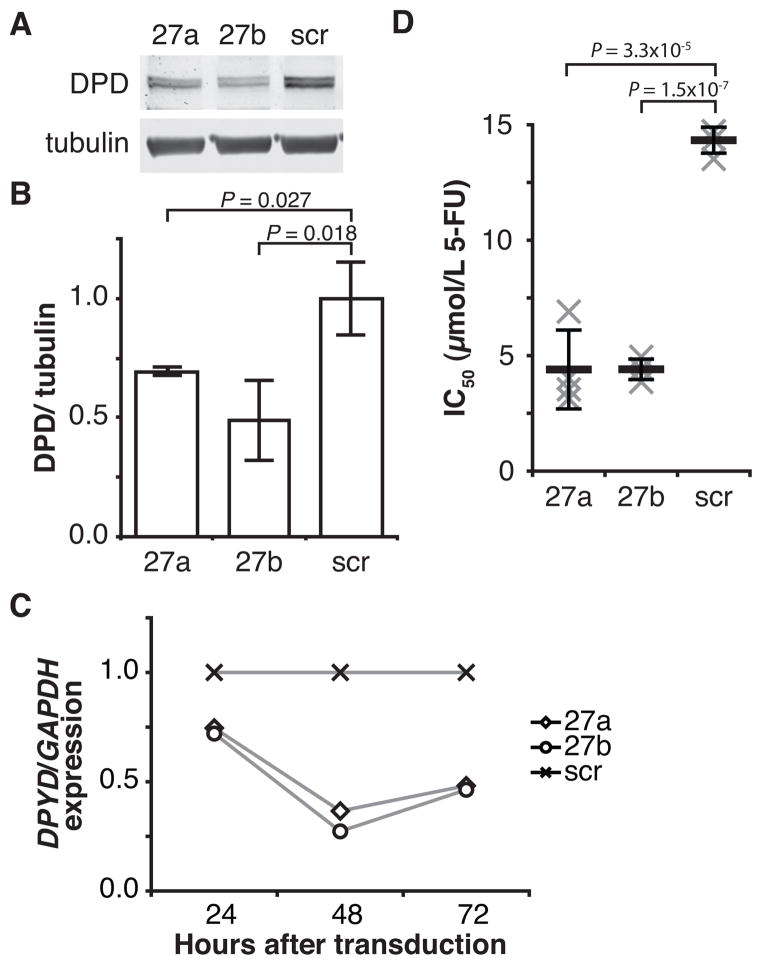
Figure 2. MiR-27a and miR-27b downregulate DPD expression and sensitize cells to 5-FU.
A, DPD and alpha-tubulin expression were measured following transduction of HCT116 cells with lentiviral particles encoding miR-27a (27a), miR-27b (27b), or a non-targeting control microRNA (scr). A representative blot is presented. B, mean DPD protein expression +/− SD for three independent replicate experiments is presented. C, expression of DPYD mRNA relative to GAPDH was measured at the indicated time points by quantitative RT-PCR. Transduction of HCT116 cells was performed as in Figure 1A. Results are presented relative to non-targeting (scr) control. A representative experiment is presented. D, the mean inhibitory concentration (IC50) for 5-FU was determined for HCT116 cells transduced as in Figure 1A. Results for each of four individual biological replicate are presented as an “x.” Mean IC50 values are represented by horizontal bars; whiskers represent standard deviations.

