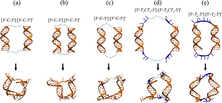
Figure 6.

Schematic representations of initial (top) and lowest SASA (below) cyclic-dimer structures after 91 ns MD simulations. Residues colored with blue are deoxythymidine (T) linkers. Note that due to the flexible T linkers, DNA deformations in (d,e) are not severe compared to (a–c) (see Table S2 in the Supporting Information).
