
Figure 1.
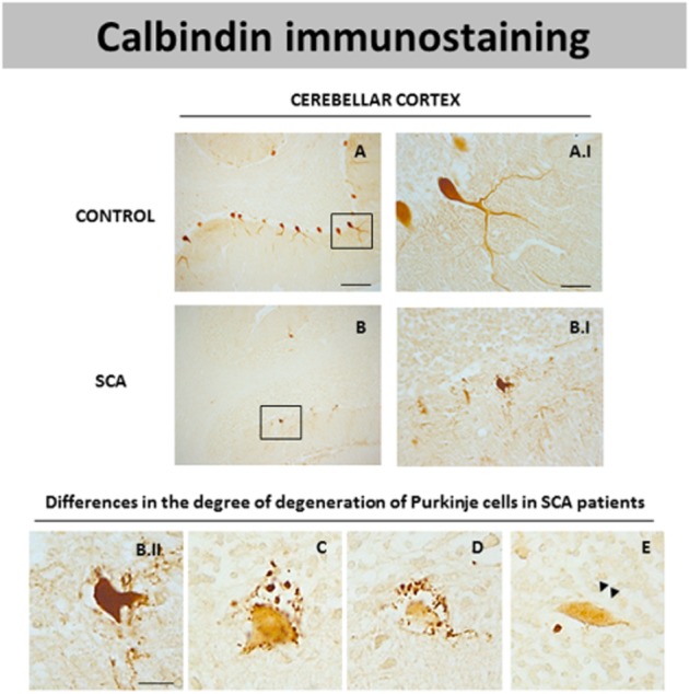
DAB immunostaining for calbindin, a marker of Purkinje cells, in the cerebellar cortex of SCA patients (B and B.I) and control subjects (A and A.I). Microphotographs shown in B.II, C, D and E correspond to details obtained in the cerebellar cortex of SCA patients proving the existence of Purkinje cells with different degrees of degeneration noted by loss in calbindin immunostaining (scale bars: A, B = 200 μm; A.I, B.I = 50 μm; B.II-E = 20 μm). Arrowheads indicate the presence of axonal torpedoes in surviving Purkinje cells.