
Figure 3.
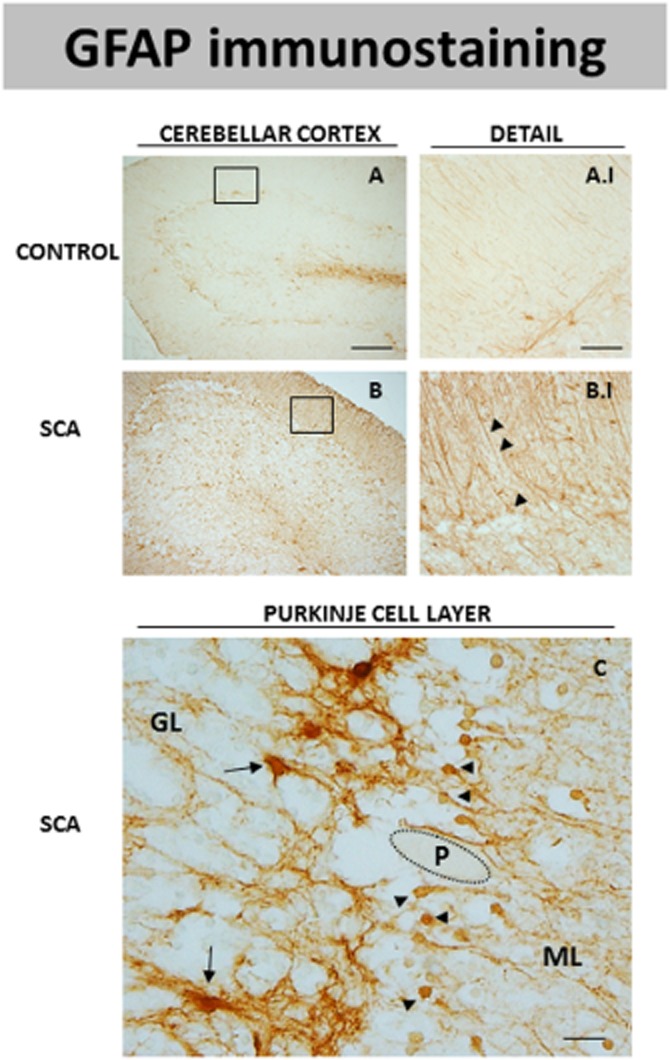
DAB immunostaining for GFAP, a marker of astrocytes, in the cerebellar cortex of SCA patients (B and B.I) and control subjects (A and A.I). The microphotograph shown in C corresponds to a detail of the Purkinje layer of SCA patients in which GFAP immunostaining was seen in two cell subpopulations that may correspond to protoplasmic astrocytes (marked with arrows) and Bergmann glia (marked with arrowheads). GL = granular layer; ML = molecular layer; P = Purkinje neurons (scale bars: A and B = 200 μm; A.I and B.I = 50 μm; C = 20 μm).