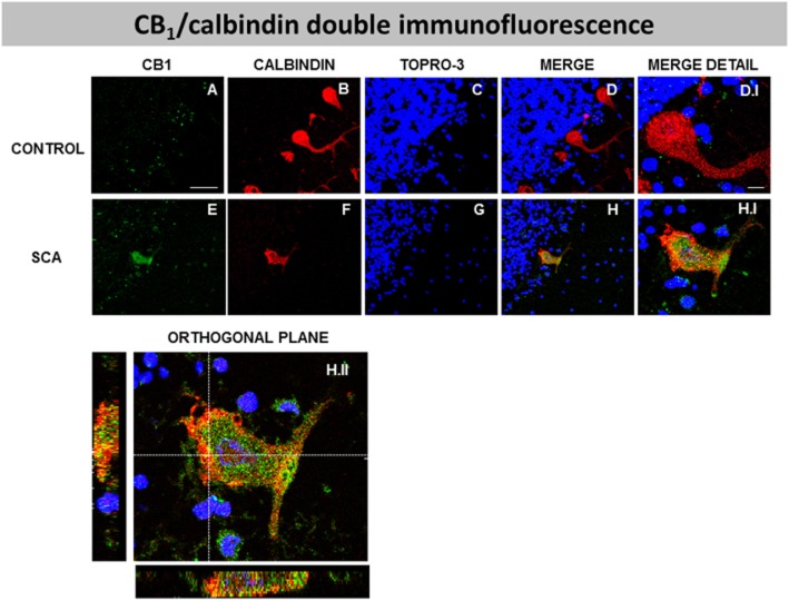
Figure 5.
Double-labelling immunofluorescence using antibodies for the CB1 receptor and calbindin, and TOPRO-3 staining, in the Purkinje layer of the cerebellum of SCA patients (E–H and H.I) and control subjects (A-D, D.I), showing co-localization of CB1 receptors and calbindin in SCA patients but not in control subjects (scale bars: A–H = 50 μm; D.I and H.I = 10 μm). The H.II panel corresponds to the orthogonal reconstruction from confocal z-series in x-z (below) and y-z (left) planes. This reconstruction confirms the co-localization of CB1 receptors and calbindin in SCA patients (scale bar: H.II = 10 μm). The microphotographs of SCA cases correspond to subject #7 (SCA2).

