Figure 7.
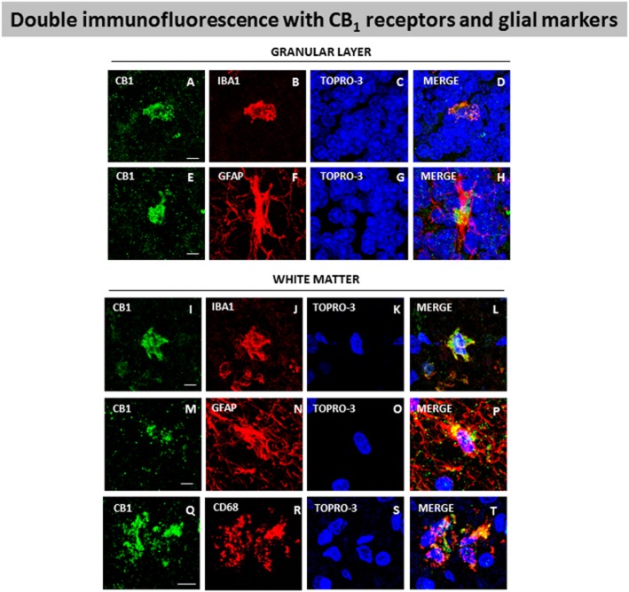
Double-labelling immunofluorescence using antibodies for the CB1 receptor and markers of glial elements (Iba-1 for microglia and GFAP for astrocytes) or infiltrated macrophages (Cd68), and TOPRO-3 staining, in the granular layer and white matter of the cerebellum of SCA patients. The microphotographs showed co-localization of CB1 receptors with Iba-1 in the granular layer (A–D) and the white matter (I–L), with GFAP in the granular layer (E–H) and the white matter (M–P), and with Cd68 in the white matter (Q–T) (scale bars: A–T = 20 μm). The microphotographs of SCA cases correspond to subject #8 (SCA), except in the case of Cd68 immunostaining that corresponds to subject #9 (SCA7).

