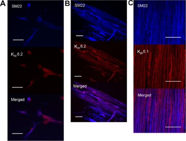
Figure 6.

Localization of KATP channels in the duodenum of rats by immunofluorescence. (A) Representative photos of the co-localization of KIR6.2 and SM22 in the isolated smooth muscle cells from duodenum. All the SM22-immunoreactive smooth muscle cells were KIR6.2-immunoreactive. (B) and (C) Representative photos of the double labelling of KIR6.2 and SM22, KIR6.1 and SM22 in the LMMP. KIR6.2 or KIR6.1 was co-expressed with SM22 in smooth muscles. Scale bars are 50 μm.
