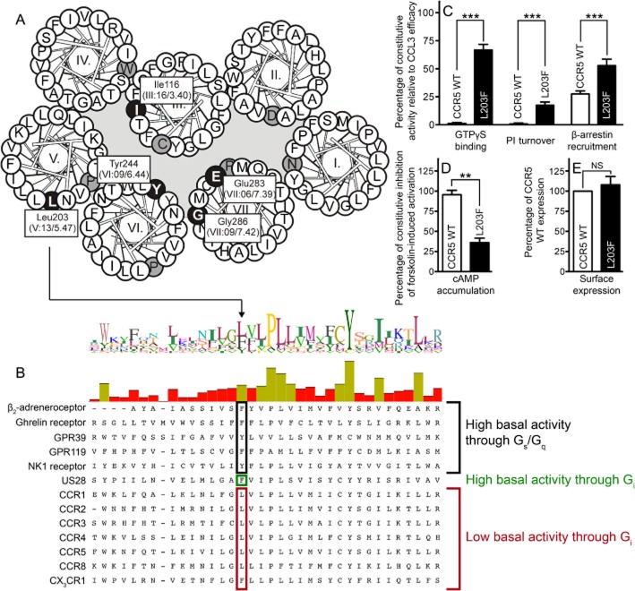
Figure 1.
Ligand-independent activation of CCR5 WT and [L203F]-CCR5. Helical wheel diagram of CCR5 indicating central residues (white on black) either mutated or of general importance for CCR5 (A). The most conserved amino acid in each TM is indicated (black on grey). (B) Alignment of class A 7TM receptors discussed in the text or similar to CCR5. Position V:13/5.47 is indicated. In addition, the level of constitutive activity in CCR5 WT and [L203F]-CCR5 in four different signalling pathways is depicted (C and D). The β-arrestin recruitment was assessed in U20S cells whereas COS-7 cells were used for the remaining. The data were normalized to CCL3 Emax on the respective receptor (C) or forskolin-induced cAMP activation in untransfected cells (shown as inhibition) (D). (E) Surface expression measured with elisa in COS-7 cells using N-terminal FLAG-tagged receptors. Data were normalized to WT. Statistical significance was calculated using Student's unpaired t-test. **P < 0.05, ***P < 0.001; ns, not significant; n = 3–25.

