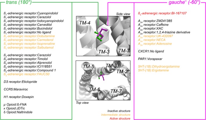
Figure 7.
Overview of the orientation of the χ1-angle of Ile in position III:16/3.40 in all relevant crystal structures (46 of the available 75 structures contain Ile in this position). The middle panels depict a side and top view the orientation of Ile in gauche− (−60°, magenta) and trans (180°, orange) position, represented by the β2-adrenoceptor structures [PDB #2RH1 (inactive) and #3SN6 (active)]. Right and left panels list the position of Ile side chain in each structure for gauche− and trans respectively. Different PDB files with identical complexes (identical ligand and receptor) are only listed once. The structures were taken from the Protein Data Bank (http://www.rcsb.org/pdb) and visualized in Molsoft Browser Pro (Molsoft LLC).

