
Figure 1.
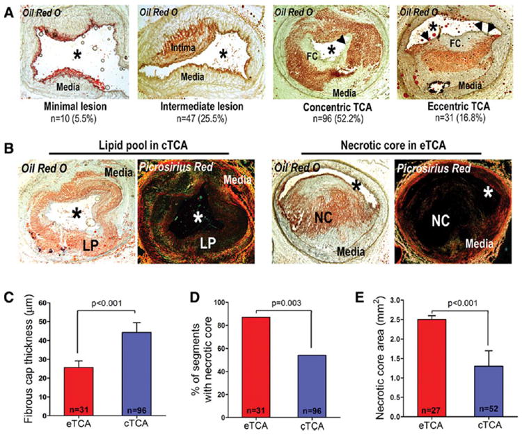
(A) Classification of segments at follow-up as segments with minimal lesion, intermediate lesion, concentric thin-capped atheroma (cTCA), and eccentric thin-capped atheroma (eTCA) plaque morphology. (B) Representative cTCA containing a collagen-positive lipid pool (LP; left) versus an eTCA containing a necrotic core (NC), i.e., lipid-rich region with absence of collagen (right). (C-E) eTCA had thinner fibrous caps, more frequently contained true NC, and had greater NC area compared to cTCA.