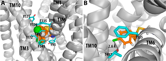
Figure 2.

Docking of compound 4h in the S1 binding site of WT SERT. Panel A is an overall view of the binding pose of compound 4h in the binding site. Panel B is a zoom-in view showing the interaction with Thr497 from TM10. The dashed line indicates favorable halogen bonding between 4h and the side chain OH group of T497 in WT SERT, while a similar interaction between 4h and A497, in the mutant, is absent, resulting in a reduction in binding affinity.
