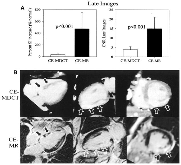
Fig. 7.
Cardiac MR compared to cardiac CT to assess for viable myocardium. Panel a The percent signal intensity (SI) and the contrast noise ratio (CNR) was higher with CE-MR (contrast-enhanced magnetic resonance image) than CEMDCT (contrast-enhanced multidetector CT). The images in panel b illustrate the same findings. Images adapted and reproduced with permission from Gerber et al. [65]

