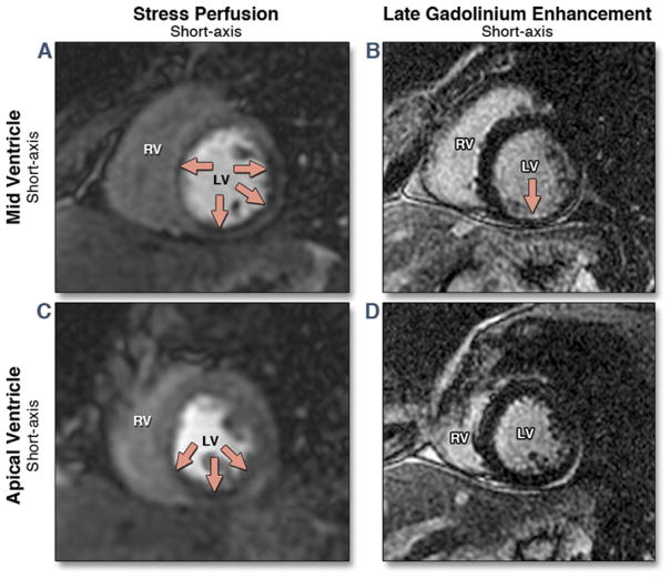
Figure 3. Stress CMR Study From a 47-Year-Old Woman With a Previous MI Referred for Assessment of Myocardial Ischemia.
Mid and apical short-axis views of the stress perfusion images show an extensive perfusion defect within the mid and apical inferior, inferoseptal, and inferolateral walls (red arrows) (A,C). Matching late gadolinium enhancement (B,D) demonstrates a small subendocardial myocardial infarction (MI) within the mid-inferior wall. CMR = cardiac magnetic resonance; LV = left ventricle; RV = right ventricle.

