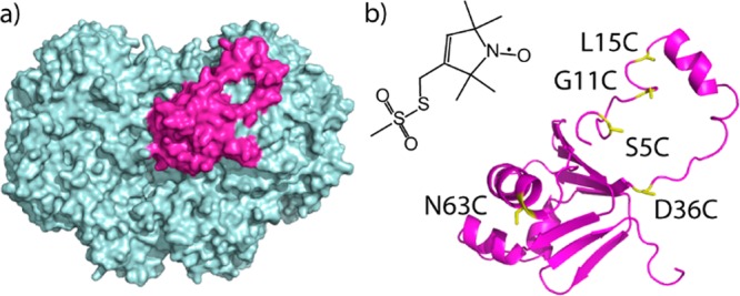
Figure 1.

X-ray crystal structure of Hox–2B complex (Protein Data Bank (PDB) ID 4GAM). (a) View of the structure, with MMOH in cyan and MMOB in magenta. There is a second MMOB bound on the other side of MMOH. (b) Structure of the spin label, MTSL. Also depicted is MMOB in the Hox–2B complex, indicating two positions (N63C and D36C) within the core and three (S5C, G11C, and L15C) on the N-terminal tail, labeled for MMOB core-to-tail distance determinations.
