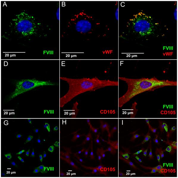
Figure 5. Immunofluorescence Analysis Demonstrates Homogeneous Staining of FVIII in MSC.
MSC and HUVEC were transduced with an expression/secretion optimized porcine FVIII in order to visualize intracellular FVIII localization. FVIII was detected by staining with an antibody against FVIII (green), while CD105 (red) staining reveals the MSC cell surface. All images were taken on a Fluoview 1000 confocal microscope with a 40x objective with various amounts of digital zoom factors as indicated by scale bars. (A-C) FVIII-transduced HUVEC co-store FVIII and VWF (red) in granules, and were used as a control. (D-F) High magnification of human MSC showing homogenous cytoplasmic staining of FVIII. (G-I) Low magnification of MSC labeled with an antibody against FVIII. Nuclei are counterstained with DAPI (blue).

