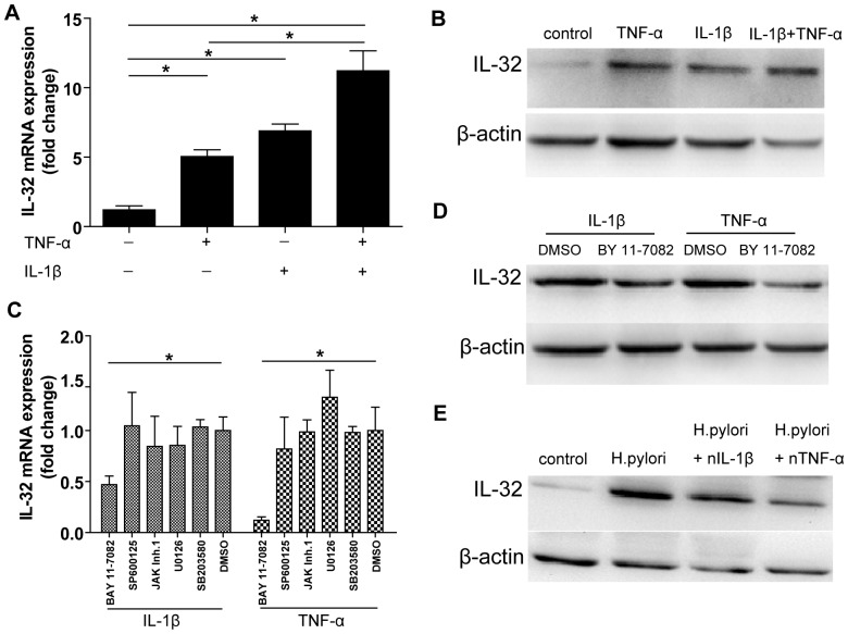
Figure 3. Induction and regulation of IL-32 mRNA in AGS cells.
A. IL-32 mRNA expression was analyzed in AGS cells after stimulation with TNF-α and/or IL-1β for 24 hours. *P<0.05; B. IL-32 protein expression was detected by Western blot from (A); C. AGS cells were pretreated with 10 µM NF-κB inhibitor (BAY 11-7082), MEK1/2 inhibitor (U0126), p38/MAPK inhibitor (SB203580), JNK inhibitor (SP600125), JAK inhibitor I or the vehicle DMSO for 1 hour prior to IL-1β or TNF-α stimulation. IL-32 mRNA level was determined by real-time PCR. *P<0.05 versus the vehicle DMSO treated cells; D. The level of IL-32 protein was detected after AGS cells pretreation with 10 µM NF-κB inhibitor (BAY 11-7082) or not and then stimulation with TNF-α or IL-1β; E. AGS cells were infected by H. pylori at a MOI = 100 for 24 hours and simultaneously treated with neutralizing antibodies to block TNF-α or IL-1β.

