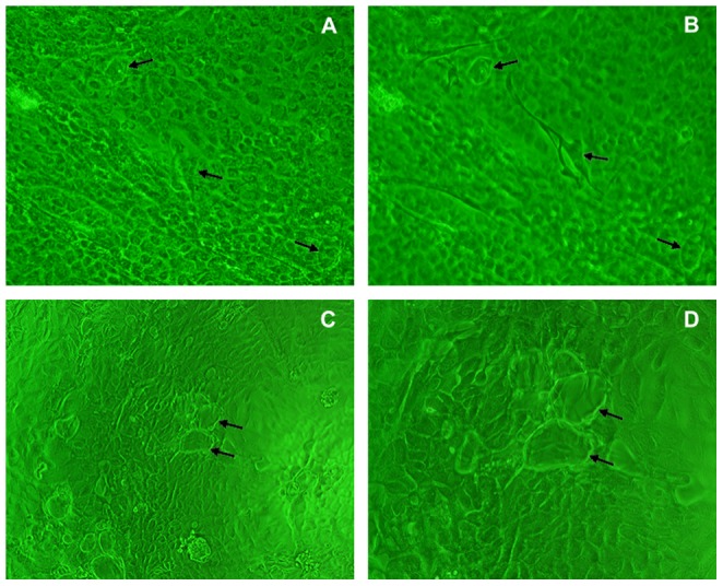
Figure 3. Developed morphology of confluent monolayer of OECs.

A: Cuticularized cells originating from post-confluent stage OECs (×200); B: Phase contrast image with the focus set on top of the raised cuticularized cells (×200); the arrows indicate typical cuticularized cells. C (×100) and D (×200): The dome structure (arrows) of the raised layer of cells above the plastic substratum.
