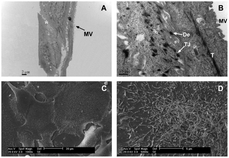
Figure 5. Transmission electron micrograph (TEM, A and B) and scanning electron micrograph (SEM, C and D) of OEC monolayers.
A: A polarized monolayer was established with apical microvilli (MV) and a basal lamina on the plastic substratum; B: Connections between neighboring cells via desmosomes (De), tight junctions (TJ) and basolateral membrane infoldings (*) were visible. Tonofilaments (T) were also observed in the OECs. C and D: SEM observations revealed numerous microvilli-like structures on the surface of the OEC monolayer.

