Figure 6. Immunofluorescence staining of OECs.

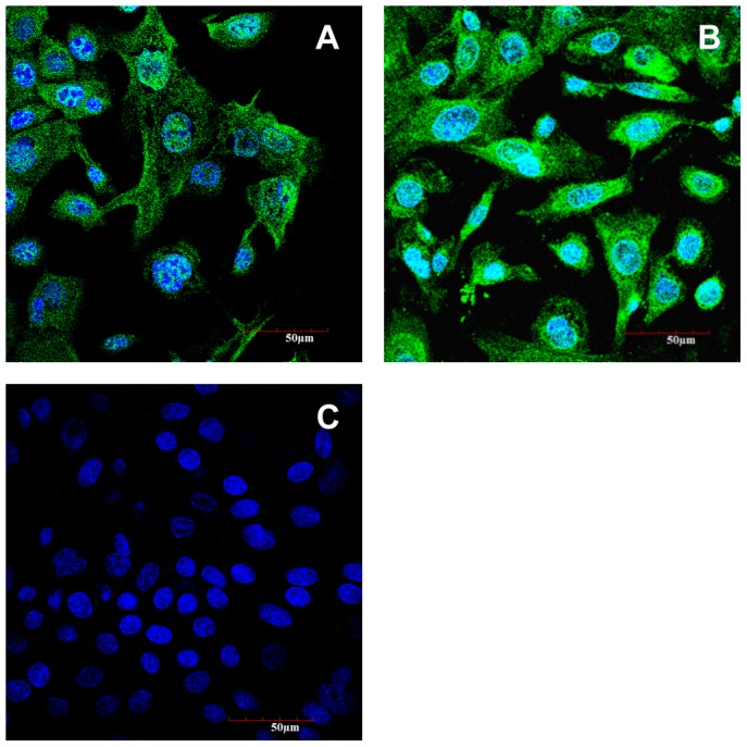
Fluorescent images of OECs stained for cytokeratin 18 (A), PEPT1 (B) and negative control using the secondary antibody only (C). Nuclei were stained with DAPI.

Fluorescent images of OECs stained for cytokeratin 18 (A), PEPT1 (B) and negative control using the secondary antibody only (C). Nuclei were stained with DAPI.