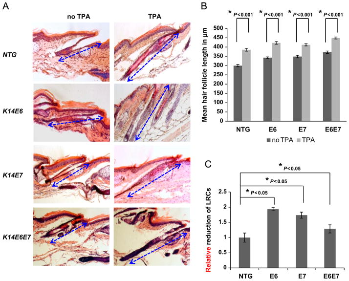
Fig. 2. Expression of the HPV16 oncogenes leads to more rapid mobilization of LRCs in response to acute anagen induction.
(A) In order to induce anagen in mice where LRCs were labeled, TPA was applied on mice every 48 h for four times. H&E staining was performed on all tail hair follicles. Hair follicle length was quantified to verify effective anagen induction. (B) ~70 Hair follicles were selected from at least 3 mice of each genotype, NTG, E6, E7 and E6E7 mice. The mean hair follicle length in μm was measured and plotted for each genotype (columns); bars, SD. All statistical comparisons were performed using a two-sided Wilcoxon rank sum test. Statistical significance was also observed between NTG and transgenic mice under resting (no TPA) conditions. (C) In order to track the mobilization of LRCs in response to acute anagen induction, the percentage reduction of LRCs was tracked per genotype. At least 3 mice of each genotype at anagen (TPA) and telogen (no TPA), NTG, E6, E7 and E6E7 mice, were selected and hair follicle bulge regions were quantified. The relative reduction of BrdUrd positive cells per hair follicle bulge was plotted for each genotype (columns); bars, SD. All statistical comparisons were performed using a two-sided Wilcoxon rank sum test. Statistical significance was also observed between E6 and E6E7 mice.

