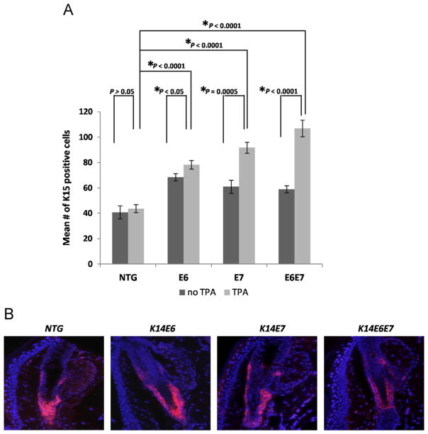
Fig. 4. Other markers of bulge stem cells are not reduced in response to HPV16 oncogene expression.
(A) Immunofluorescence was performed using a K15-specific antibody. ~50 Hair follicles were selected from at least 3 mice of each genotype, NTG, E6, E7 and E6E7 mice. The mean number of K15 positive cells of each hair follicle was quantified and plotted for each genotype (columns); bars, SD. All statistical comparisons were performed using a two-sided Wilcoxon rank sum test. Statistical significance was also observed between the NTG and the transgenic mice (no TPA) as well as between the various transgenics (TPA-treated). (B) Representative immunofluorescence of K15 staining (red) in the hair follicles of the genotypes examined. Counterstaining was done with DAPI (blue).

